
 Deutsch
Industry-leading eye laser technology worldwide
We use the best available technology and methods worldwide.
Free info kit
Free quick check


We use the best available technology and methods worldwide.
Free info kit
Advances in laser eye technology have revolutionized the capabilities of refractive surgery, leading to significantly improved laser eye surgery outcomes for patients. These technologies enable more precise corrections of vision defects such as nearsightedness, farsightedness, astigmatism, and presbyopia, resulting in higher patient satisfaction.
One of the key aspects of laser eye technology is its ability to shape the cornea of the eye with extraordinary precision. Procedures like FemtoLASIK use a highly precise laser to cut and reshape the cornea, leading to correct light refraction and thus improved vision quality. Modern methods like SMILE Pro even use a femtosecond laser, which further increases precision and minimizes the risk of complications.
In addition to precision, LASIK technologies also offer quick recovery times. Many patients report a significant improvement in their vision shortly after the procedure. This is a considerable advantage over traditional methods, where recovery times can be longer.
The continuous development and improvement of laser eye technologies contribute to making refractive surgery safer, more effective, and more accessible than ever before. For people affected by vision defects, this offers a promising opportunity to improve their vision and potentially reduce or even eliminate dependence on glasses or contact lenses.
In summary, the importance of laser eye technology for refractive surgery cannot be overstated. It offers patients not only the prospect of better vision quality but also faster recovery and a significant improvement in their quality of life.
As the Lasikwelt Laser Eye Center Cologne, we are committed to offering the best available technology and methods worldwide.

With a comprehensive, well-thought-out concept, ZEISS has assembled an optimally matched refractive system landscape, consisting of the MEL® 90 Excimer Laser, the VisuMax® 800, and the CRS-Master® treatment planning station. When used simultaneously, these three premium devices ensure a streamlined workflow and effective patient care.
Refractive surgery has rapidly evolved over the past decades, enabling more precise correction of vision defects than ever before. A critical factor for the success of such procedures is careful planning and precise execution. This is where the ZEISS Refractive Workplace comes in, an innovative platform that optimizes and supports the entire process of refractive surgery.
The ZEISS Refractive Workplace is a comprehensive software solution developed by the renowned company Carl Zeiss Meditec. It serves as an interface between diagnosis, surgical planning, and execution. With its help, ophthalmologists can integrate patient-specific data from various sources, simulate and optimize treatment strategies, and precisely configure the operational settings for refractive laser devices.
The core of the ZEISS Refractive Workplace is a user-friendly software that visualizes all relevant patient data. It enables detailed analysis of the front and back surfaces of the cornea, wavefront data, and lens density. Doctors can use it to create individual treatment plans that consider both the anatomical peculiarities and the specific visual requirements of the patient.
One of the greatest advantages of the ZEISS Refractive Workplace is the ability to standardize surgical processes while ensuring individualized treatment. The intuitive user interface and integration of data from various measuring devices increase efficiency in daily clinical practice and help improve the safety and predictability of procedures.
The ZEISS Refractive Workplace is used in the planning and execution of various refractive procedures, from LASIK to PRK to lens implants. It is especially valuable when applying complex technologies like SMILE (Small Incision Lenticule Extraction) or toric intraocular lenses, where precise measurements and positioning are crucial for success.
The future of refractive surgery lies in precise customization for each patient. The ZEISS Refractive Workplace forms a solid foundation for even more personalized patient care and surgical excellence. With ongoing development and integration of further innovative technologies, this platform will continue to enhance the quality and outcomes of refractive procedures.
The ZEISS Refractive Workplace exemplifies technological progress in ophthalmology. It combines diagnostics and surgical planning in a seamless process that allows doctors to work at the highest level and provide patients with the best possible vision.
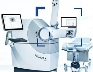 The Carl Zeiss Meditec VisuMax® 800 Femtosecond Laser is the latest in the third generation of femtosecond lasers. It is more precise and energy-efficient than any other laser system. The VisuMax is so accurate that it uses up to seven times less energy than other femtosecond lasers. Low energy consumption is crucial when treating the delicate tissue of the cornea. The high-precision optics from ZEISS offer an extremely fixed laser beam that achieves the desired ablation depth with minimal laser energy, leaving the tissue outside the targeted area untouched. The result is minimal and thus more precise laser energy to create more accurate, sensitive, and gentler flaps than any other femtosecond laser.
The VisuMax® 800 is thus the only femtosecond laser that does not increase intraocular pressure, so you will not feel anything during the treatment. Due to its unique accuracy, the VisuMax is the only femtosecond laser suitable for the most advanced laser eye treatment method: Smile PRO, ReLEx-SMILE.
The Carl Zeiss Meditec MEL® 90 Excimer Laser
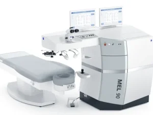
The Excimer Laser – The Intelligent Connection of Proven Experience and Exciting Progress. The use of excimer laser technology in refractive treatments began in 1986 with Zeiss. The MEL® 90 continues this tradition. A successful combination of expertise and innovation promises the user a rewarding, comprehensive experience.
The Zeiss MEL® 90 Excimer Laser offers incredibly precise, self-stabilized, calibrated laser energy. Zeiss was the first laser manufacturer to produce so-called close-loop energy stabilization – the common method of energy stabilization used by the MEL® 90. This gives it the fastest response time of all lasers without the constant need for user calibration.
The MEL 90 is also a top-notch program for treating patients with presbyopia. ZEISS optionally offers the advanced and genuinely binocular PRESBYOND® Laser Blended Vision module, which far exceeds the limitations of conventional monovision.
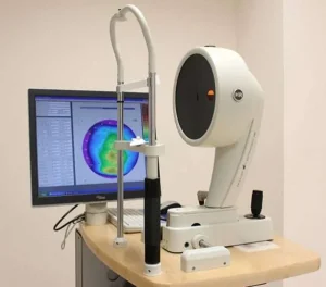 Pentacam HR: Revolution in Corneal Diagnostics
In ophthalmology, a precise examination of the cornea is crucial for diagnosing and treating vision defects and corneal diseases. The Pentacam HR represents a revolutionary advancement in corneal measurement and offers ophthalmologists a comprehensive view of the anterior eye segments.
The Pentacam HR is a high-resolution Scheimpflug camera system that provides a detailed depiction and analysis of the cornea. The device is characterized by its ability to capture thousands of measurement points on the cornea to create a three-dimensional model of the anterior eye segment. This highly precise technology allows for a comprehensive analysis of the cornea, including the front and back surfaces and corneal thickness.
Using a rotating Scheimpflug camera, the Pentacam HR performs a 360-degree scan of the cornea, generating images in seconds. These images are processed into a three-dimensional model that allows for exact measurement and visualization of the corneal structures. Through the three-dimensional analysis, subtle changes and anomalies can be detected that would otherwise remain invisible to the human eye.
The Pentacam HR offers significant advantages over other diagnostic methods. It not only enables a contactless and patient-friendly examination but also provides extremely accurate data on corneal topography and tomography. These data are especially important before refractive surgery, in diagnosing keratoconus and other corneal dystrophies, and in cataract and glaucoma assessment.
The device is used in routine diagnostics and specialized eye centers. It is an indispensable tool for planning laser eye treatments, selecting and fitting intraocular lenses, and fitting contact lenses. It is also essential for early detection and management of corneal diseases.
The Pentacam HR opens new possibilities in personalized medicine. By integrating artificial intelligence and machine learning into the evaluation software, diagnoses can be even more precise and treatment recommendations even more personalized in the future. This contributes to a continuous improvement in patient care and cements the Pentacam HR’s role as the gold standard in corneal diagnostics.
With the Pentacam HR, ophthalmologists have an advanced device at their disposal that takes precision in corneal diagnostics to a new level and opens the door to a new era in ophthalmology.
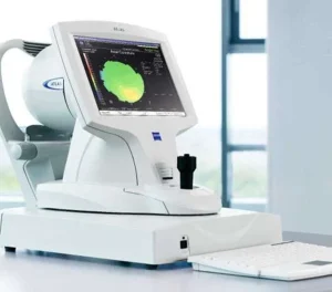
The ATLAS 9000 corneal topography system, utilizing Placido Disk technology with the Cone-of-Focus™ alignment system, offers an innovative and precise solution for corneal topographic analysis. The unique combination of the proven Cone-of-Focus™ alignment system and the sophisticated Arc-Step algorithm allows for sub-micrometer precision measurements and reliable surface reconstruction through triangulation.
The enhanced effectiveness of the topography system is derived from a 22-ring Placido Disk system, which avoids the overlap of projection rings and ensures accurate measurements for a wide range of patients. Additionally, the generous working distance of 70 mm reduces measurement errors, which are more common with systems with smaller cones.
With SmartCapture™ image analysis technology, the ideal image is captured during the first measurement. Up to 15 digital images are taken per second during alignment, and the best one is automatically selected. The next generation of image processing ensures repeatable and reliable results, even in complex cases.
The advanced corneal wavefront analysis improves patient consultation and optimizes treatment selection. Using ray-tracing technology, higher-order corneal aberrations are made visible, providing essential insights for consultation and treatment planning.
This method of analysis offers additional benefits, including:
Furthermore, automatic pupillometry and HVID measurement facilitate contact lens fitting and planning of refractive procedures. The pupil size, measured under two lighting conditions, provides important information about the optical zone under various lighting conditions. This procedure helps assess the centering and effectiveness of LASIK or orthokeratology and supports the fitting of multifocal contact lenses by providing a better understanding of pupil size. Additionally, the selection and fitting of contact lenses are optimized through the automatic measurement of the Horizontal Visible Iris Diameter (HVID).
The ergonomically designed system is ideal for surgeons and patients:
It integrates a powerful computer with advanced analysis software in a compact design. The innovative chin rest allows for optimal positioning of patients, facilitates image capture, covers a wide range of the corneal periphery, and automatically recognizes the left and right eyes. The invisible lighting of the Placido rings ensures comfort even for light-sensitive patients.
Maximize the efficiency of your topographic analysis.
With PathFinder II software for the ATLAS corneal topography system (model 9000), topographic screenings before laser eye treatments can be conducted easily and safely. The software supports screening for refractive surgery and helps identify abnormal corneal conditions. Advanced analysis methods of PathFinder II provide immediate risk assessments for various corneal changes based on a wide range of topography data and a comprehensive clinical database.
For perfect fitting of gas-permeable lenses.
Complex corneal conditions such as asymmetric astigmatism or irregular astigmatism affect contact lens fit. The MasterFit II software allows for customized GP lens fitting according to your needs and offers numerous benefits:
Fluorescein image simulation supports the fitting of specialty and stock lenses, including spherical and toric variants. The software automatically adjusts the lenses to ensure optimal tear film distance, improving efficiency in lens fitting by adjusting parameters and simulating lens movement. The transfer of topography results to your lab is done electronically to simplify ordering and fitting.
Improve your productivity with connected solutions.
Atlas Review software is designed specifically for users of models 993, 995, and 9000, building on the proven features of previous versions. With this tool, you can significantly expand the capabilities of your existing Atlas corneal topography system:
Thanks to dynamic remote access to all topographic examination data and patient education tools, such as corneal wavefront simulation, you can accelerate your workflows. The software offers similar functionality to the Atlas model 9000 and is compatible with earlier models 993 and 995.
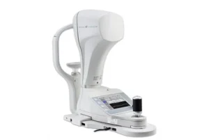 The Corvis® ST – Insight into Modern Diagnostics of Corneal Biomechanics
In the world of ophthalmology, corneal biomechanics play a central role in assessing eye health and planning surgical procedures. With the Corvis® ST, a highly advanced diagnostic device is available, setting new standards in this area.
The Corvis® ST is a non-invasive tonometric device equipped with an ultra-high-speed camera to capture the dynamic response of the cornea to a standardized air impulse. This technology allows precise measurements of the biomechanical properties of the cornea, providing a valuable complement to traditional methods of corneal measurement.
The principle of the Corvis® ST is based on a controlled air puff that deforms the cornea. During this process, the high-speed camera, capturing over 4,300 images per second, records changes in the corneal structure. The resulting data provide information not only about intraocular pressure but also about the biomechanical properties of the cornea, such as its elasticity and strength.
The data provided by the Corvis® ST enable a comprehensive analysis of corneal biomechanics. This allows for the early identification of patients at risk for diseases such as keratoconus. Patients considering refractive surgery also benefit, as the Corvis® ST provides important information about the stability and resilience of the cornea.
Additionally, the Corvis® ST contributes to precise diagnosis and monitoring of glaucoma. By measuring the cornea’s biomechanical behavior, more accurate statements about the actual intraocular pressure, a crucial factor in glaucoma, can be made.
In addition to glaucoma diagnostics and pre-refractive surgery evaluation, the Corvis® ST is used in postoperative follow-up and corneal biomechanics research. Researchers and clinicians appreciate the device for its ability to detect subtle changes in the cornea that may go unnoticed with traditional methods.
With its advanced technology and ability to capture complex biomechanical data, the Corvis® ST stands at the forefront of diagnostic innovations in ophthalmology. The continuous development and integration of device data into large-scale clinical databases promise to deepen the understanding of corneal diseases and further improve treatment outcomes.
The Corvis® ST not only represents the current state of the art but also points to the direction in which ophthalmology will move in the future – towards more precise, personalized patient care

WaveScan technology allows advanced wavefront analysis to identify vision defects that go beyond what can be corrected with traditional vision aids. This method captures not only standard refractive errors but also more complex higher-order aberrations. Such precise data are essential to enable customized laser treatment that optimally improves the patient’s vision.
During the WaveScan analysis, 168 specific points on the cornea are examined to determine the refractive properties of the eye. This includes measuring 24 optical irregularities of higher order in addition to the usual refractive errors such as myopia, hyperopia, and astigmatism, which are incorporated into the laser correction.
In the wavefront measurement, the device sends light pulses through the eye to the retina. The reflected waves are sent back through the cornea and lens and captured by a wavefront sensor. A so-called fundus camera images these waves, making the distortions of the outgoing rays visible and allowing the errors in the entire optical system of the eye to be determined.
After the wavefront measurements are completed, our surgeons have detailed information about the patient’s individual vision defects. Based on these data, the most suitable laser eye treatment method, whether ReLEx SMILE / SMILE Pro, LASIK / FemtoLASIK, or PRESBYOND, is selected for the individual patient.
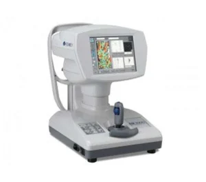
The Endothelial Microscope EM-3000 represents a significant advancement in the technology used for assessing corneal endothelial cells. This cutting-edge device enables precise measurement of the cell density and morphology of the cornea’s innermost layer, all without any physical contact with the eye. This non-invasive approach is critical for ensuring patient comfort and safety during the examination process.
The EM-3000 features the capability to select from seven different imaging modes, allowing for a tailored approach depending on the specific requirements of the examination. Within a very short period, the microscope can capture up to 15 images per series, ensuring that the best possible image is automatically highlighted for detailed analysis. This high level of imaging efficiency and accuracy is essential for thorough corneal assessment.
It is strongly recommended to perform annual checks of endothelial cells using specular microscopy after the implantation of a phakic intraocular lens (PIOL), such as an Implantable Collamer Lens (ICL). Regular monitoring is vital because the integrity of endothelial cells is crucial for maintaining corneal clarity and overall eye health. The endothelial cells are responsible for continuously pumping out excess moisture from the cornea, thereby preventing corneal edema and maintaining transparency.
Moreover, this method of specular microscopy is invaluable for diagnosing conditions such as Fuchs’ dystrophy, a progressive eye disease that leads to the gradual loss of endothelial cells. Early and accurate detection of Fuchs’ dystrophy allows for timely intervention and management, potentially preserving vision and preventing severe complications.
The EM-3000’s advanced imaging technology provides detailed insights into the health and function of endothelial cells, facilitating better diagnostic accuracy and improved patient outcomes. Its ability to offer non-contact, high-resolution imaging makes it an indispensable tool in modern ophthalmology, particularly for post-operative care and ongoing monitoring of patients with phakic intraocular lenses. The device’s efficiency and precision are essential for detecting even subtle changes in endothelial cell density and morphology, ensuring that any potential issues are identified and addressed promptly.
The NIDEK Tonoref II is a multifunctional 3-in-1 device that combines an autokeratometer, an autorefractometer, and a non-contact tonometer. This combination allows for three different measurements to be conducted quickly, conveniently, and automatically in succession without having to operate different devices. Measurements include refractive value, intraocular pressure, and corneal curvature.
Even in challenging eyelid conditions, the operation of the Tonoref II is straightforward. Integrated functions ensure automatic centering of the pupil and triggering of the measurement. Additionally, the device provides instant verification of each measurement and allows for easy data transfer to various devices, including a printer.
Special Features:
A device with four measurement functions – a combination that saves both time and space. Automated focusing, centering, and triggering of measurements facilitate pachymetry, non-contact tonometry, keratometry, and refraction in direct succession.
Thanks to this innovative instrument, our surgeons can obtain extensive information about your vision and the general condition of your eyes, all during an examination where your eyes are not touched.
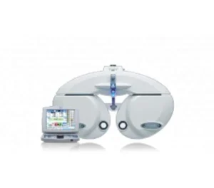 The NIDEK RT-5100 Phoropter enables precise automatic refraction measurements and is characterized by its user-friendly operation. Control is realized via a clearly structured control module with a built-in touchscreen. All vision tests of the CP-770, CP-690, and CP-660 projectors can be selected directly through this module. Additionally, infrared technology allows for controlling the SC-1600 Pola visual test screen device. Quick transfer of refraction and eyewear values is ensured through compatibility with NIDEK autorefractors of the AR and ARK series as well as the LM lens meters.
The NIDEK RT-5100 Phoropter is designed for precise and automated refraction determination, delivering exact values independent of the operator. Optimal data transfer compatibility with various devices is guaranteed. It ensures accurate measurement results under realistic viewing conditions for determining correct eyewear values. The phoropter is equipped with a special function for checking near vision. Thanks to a special coating that keeps the lenses clean, the test results are even more precise.
The phrase “Your vision performance is our primary focus” means that every patient at the LASIKWELT Laser Eye Center will receive the best eye care. Our surgeons and staff ensure the best care during your laser eye surgery. To provide the best possible eye care and patient care, we have invested in the most modern laser eye technology. With well-trained personnel equipped with the best tools in the industry, we can achieve our high goal of offering excellent care at very affordable prices and with financing options.
The next stage of development in modern laser eye surgery: Smile PRO with the Visumax 800 Femtosecond Laser. The laser eye technology at the LASIKWELT Laser Eye Center in Cologne is safer than ever before and can be tailored to the personal vision needs of the patient.
Our industry-leading and currently fastest refractive platform from ZEISS combines for the first time the two laser systems Visumax 800 Femtosecond Laser and MEL 90 Excimer Laser.
LASIKWELT Laser Eye Center Cologne


